Seeing More Clearly Now with New Research
Congratulations to Dr Inaki-Carril Mundinano, William Kwan and Professor James Bourne on their recent publication “Retinotopic specializations of cortical and thalamic inputs to area MT” in high-impact journal PNAS (The Proceedings of the National Academy of Sciences).
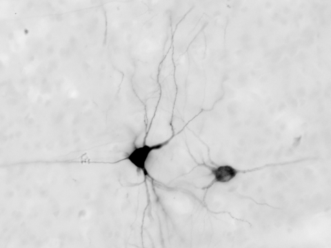
Using advanced imaging techniques, the research team investigated the anatomy, morphology and distribution of specialised neurons responsible for visual motion perception in a previously understudied part of the brain in primates. These exciting new findings change our established understanding of the anatomic organisation of the visual system.
“Having a greater grasp of how these parts of the brain develop and function is critical if we are to prevent or develop treatments for diseases or disorder were we lose the capability to process this type of information.” – Dr Inaki-Carril Mundinano
“It is well known that neurons in a certain part of the brain called the ventral visual system have special characteristics (retinotopic specialisations) that give primates and humans very accurate vision. However, these specialisations have not yet been studied in another part of the brain we were interested in, the dorsal visual stream,” said lead author Dr Mundinano.
Dr Mundinano added, “The dorsal visual stream is dedicated to processing visual motion and visually-guided behaviours, like watching players run back and forth in a football game or guiding our hands to type the right letters. So the dorsal visual stream actually plays a major part in many of our lives. Having a greater grasp of how these parts of the brain develop and function is critical if we are to prevent or develop treatments for diseases or disorder were we lose the capability to process this type of information.”
In this new publication, the research team describe a heterogeneous population and distribution of a specific subset of neurons, called V1 (visual cortex)-MT (middle temporal) projecting neurons in primates. This primate adaptation of the V1 to MT pathway is arranged in a way that we had not previously understood, with a morphology and distribution pattern that suggests a rapid relay of motion information.
“With these results, we are one step closer to fully understanding how human sight develops, how this can break down and be affected, and how we can fix it.” – Professor James Bourne
The paper also provides new evidence that supports the hypothesis that another group of neurons in the thalamus (pulvinar nucleus) is important during development, and the establishment of the specific cellular organisation observed in the area MT.
“Studying the morphology, distribution and organisation of specialised neurons give powerful clues as to how neurons function and interact with one another in a dense, delicate, interconnected and complex system,” commented Professor Bourne. “With these results, we are one step closer to fully understanding how human sight develops, how this can break down and be affected, and how we can fix it.”
Once again, congratulations to the research team!
More information
Click here to read the publication: Mundinano, I. C., Kwan, W. C., & Bourne, J. A. (2019). Retinotopic specializations of cortical and thalamic inputs to area MT. Proceedings of the National Academy of Sciences, 201909799.
The Bourne Group is at the forefront of understanding visual brain development and plasticity, as well as studying pathology states such as stroke. For more information on Professor James Bourne and his group at ARMI, please visit the Bourne Group page. You can contact Professor James Bourne via james.bourne@monash.edu.